• Positive control: ImmunoCultTM Human CD3/CD28 Activator (STEMCELL)
2
Principle
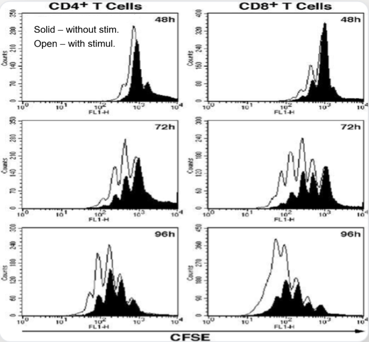
• CFSE flow cytometry tracks dye dilution during cell division to distinguish proliferation generations. Combined with Ki-67 staining, it accurately reflects T cell proliferation activity.
3
Application
• Vaccine R&D: Immunogenicity
• Tumor Immunotherapy: TIL/TCR-T
• Autoimmune: Pathogenesis
02. T Cell Activation
1
Information
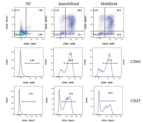
• Core Biomarkers: dual marker of CD69 and CD25
• Positive control: PMA + Ionomycin (cocktail)
2
Principle
• Quantify early stimulation marker CD69 and mid-term marker CD25 expression via flow cytometry to distinguish activated T cell proportion for evaluating immune function status.
3
Application
• Drug R&D Screening
• Basic immunology research
• Immunotherapy: CAR-T evaluation
03. Cytokine Secretion Profile
1
Information
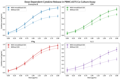
• Core Biomarkers: focus on key effectors IL-2, IFN-γ, TNF-α to reflect immune activation
• Positive control: PMA + Ionomycin (cocktail) as gold standard
2
Principle
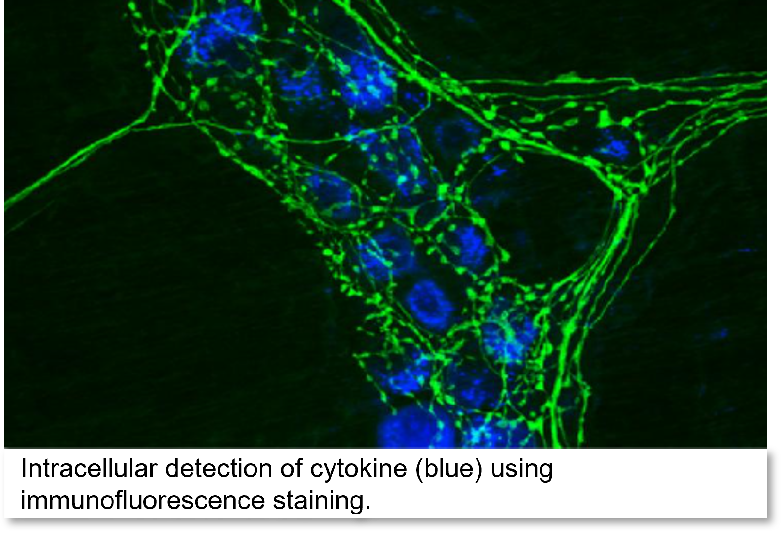
• Dual detection of cytokines in supernatant (ELISA/MSD) and single cell level (i.e. intracellular staining).
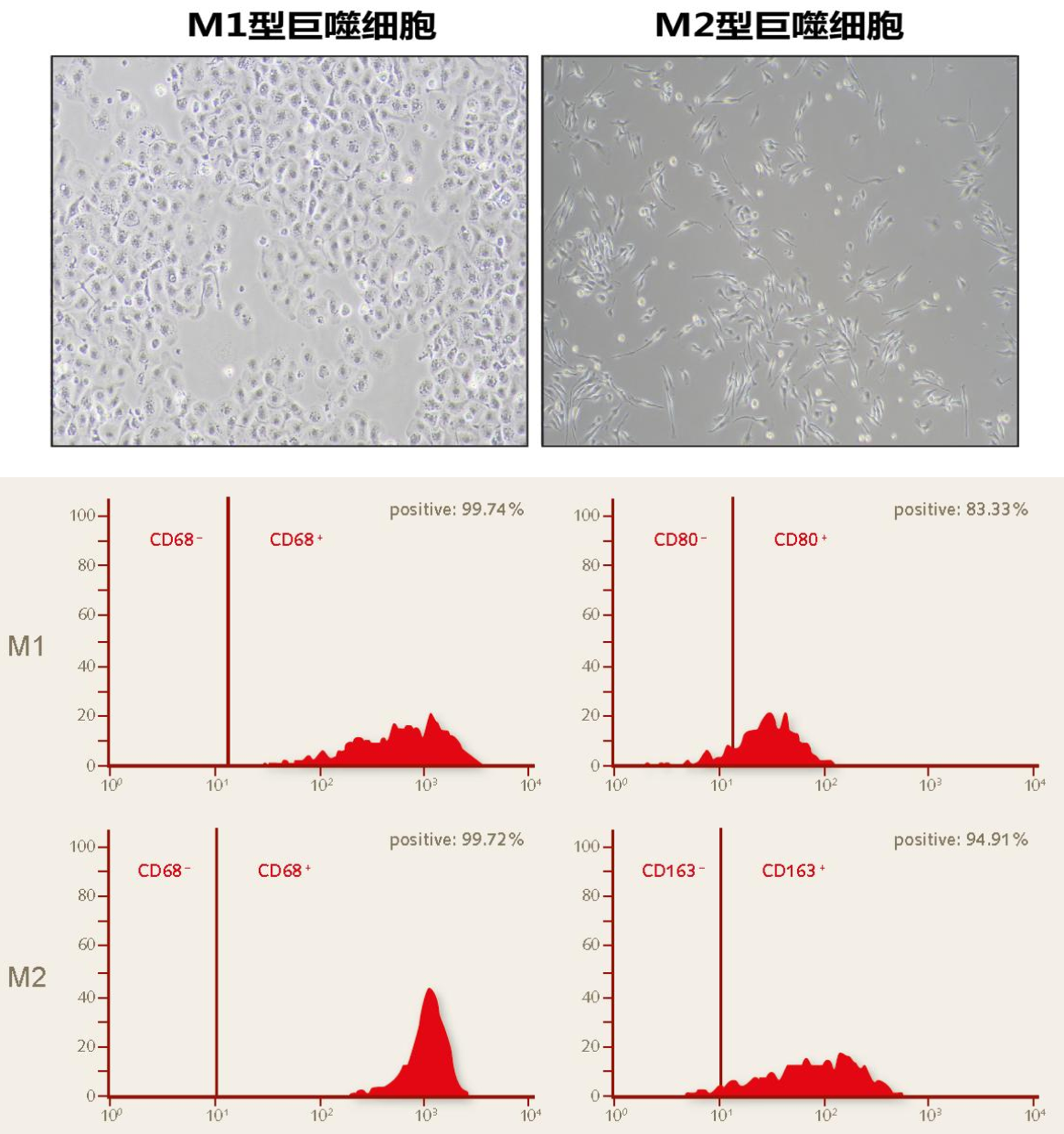
• Macrophage polarize to pro-inflammatory M1 or anti-inflammatory M2 phenotype, which can be distinguished by flow cytometry along with characteristic cytokine secretion.
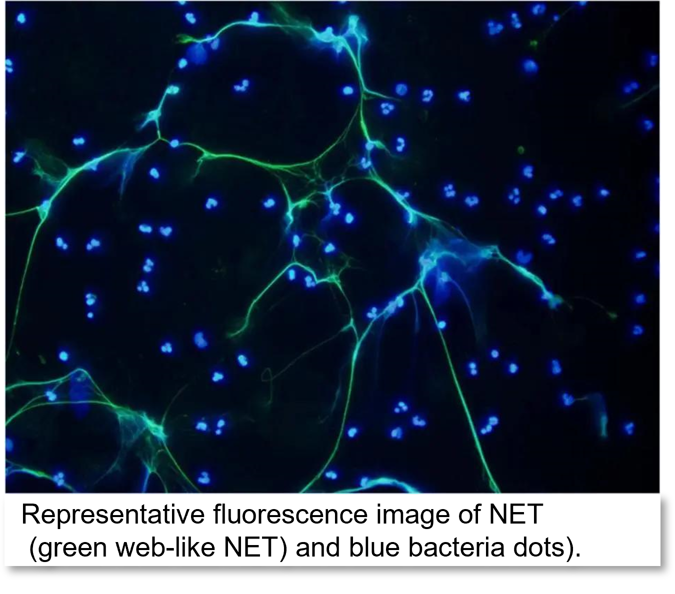
• Activated neutrophile releases Neutrophil Extracellular Trap to detain pathogens. NET is composed of DNA backbone and antimicrobial proteins (e.g. histone, myeloperoxidase, and neutrophile elastase. This sticky web immobilizes and kills invading pathogens.
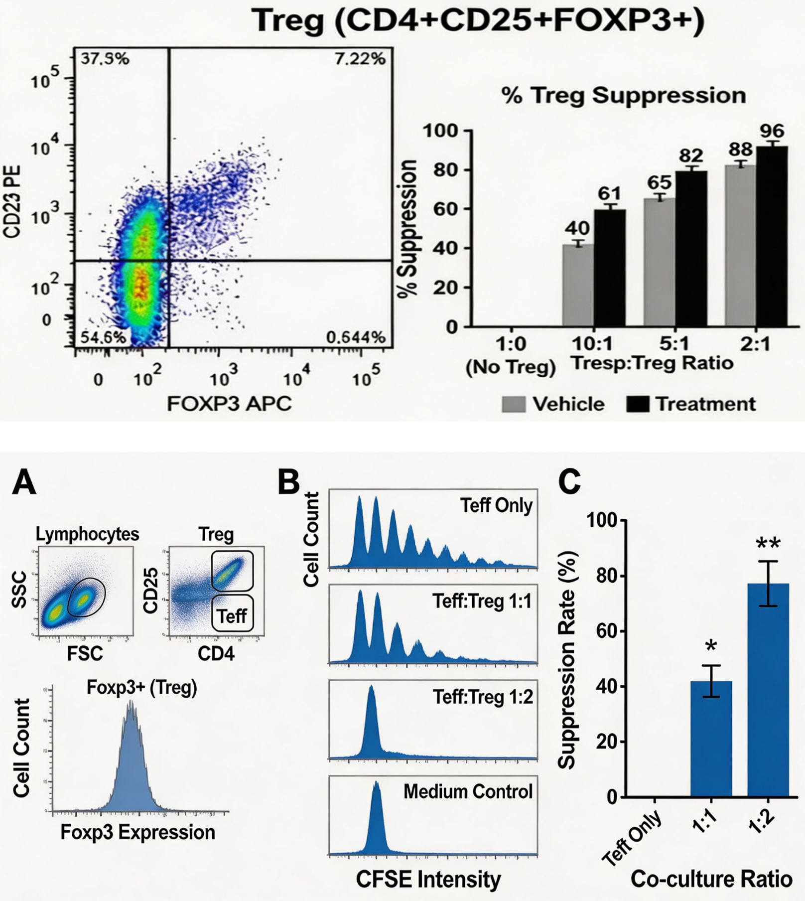
• T-regs are specialized subset of T cells that function as immune “brakes”, preventing excessive immune responses. T-regs suppress immune functions of T, B, and antigen-presenting cells (APC) by direct cell-to-cell contact and release inhibitory molecules IL-10 and TGF-β.
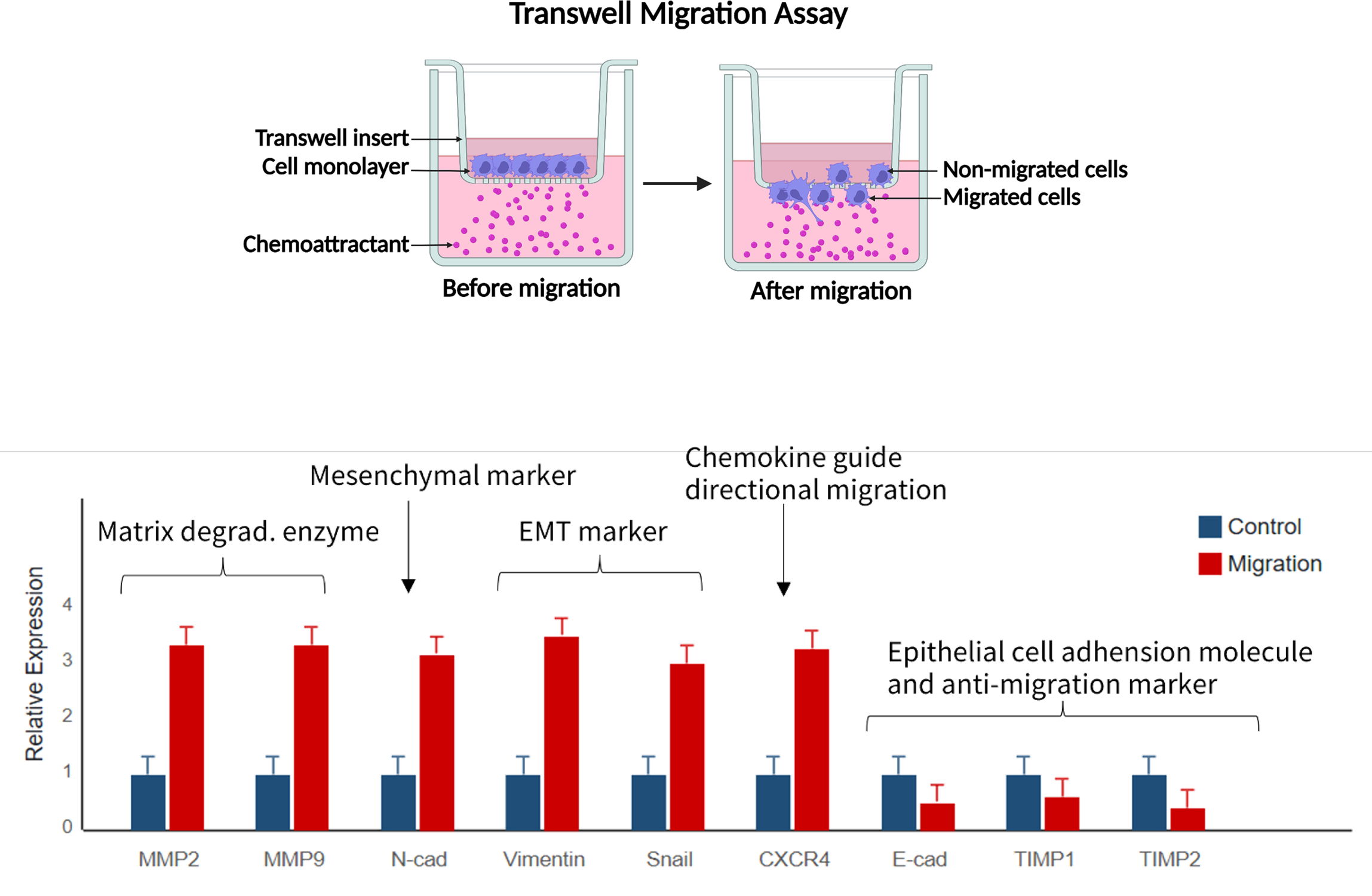
• Simulate the in vivo environment via Transwell assay to quantify immune cells’ directional migration to chemokines, and analyze chemokine receptor (e.g. CXCR3) by flow.